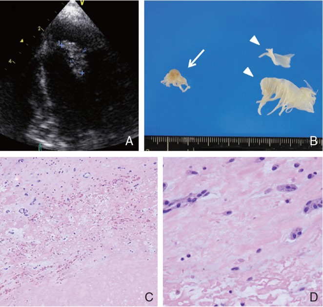
Fig. 4.

TEE visualizing a mass lesion (15 × 6 mm) adherent to the anterior cusp of the mitral valve indicating left atrial myxoma (A). Macroscopic findings of cardiac tumor (14 × 8 mm) removed by cardiosurgery (arrow), and normal anterior cusp of mitral valve (arrowhead) removed by open heart surgery (B). Specimen of cardiac tumor demonstrates paucicellular mesenchymal tumor with spindle cells in an abundant myxoid matrix, cardiac myxoma (C: H&E 40×, D: H&E 400×). TEE: transesophageal echocardiography, H&E: hematoxylin and eosin.
