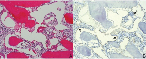
Fig. 2.
Photomicrograph of hematoxylin and eosin staining of the resected specimen revealing both bone lamella and irregular fibrous tissue, and in part, granulation tissue with neutrophils, plasma cells and lymphocytes in the marrow cavity. Original magnification ×25 (A). Immunohistochemical staining for D2-40 revealing positive endothelial cells in many dilated, thin-walled and vascular channels, as shown in light brown (solid arrows). Original magnification ×25 (B).

