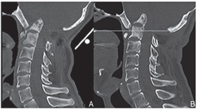
Fig. 4.

Two years after first surgery, reconstructed sagittal CT image revealing the progress of basilar impression with osteolysis of the anterior arch of C1 and isolated fragment of the odontoid process (A). At 4.2-year follow-up, CT image revealing the tip of the odontoid process more than 30 mm above McGregor’s line (dotted line) (B). CT: computed tomography.
