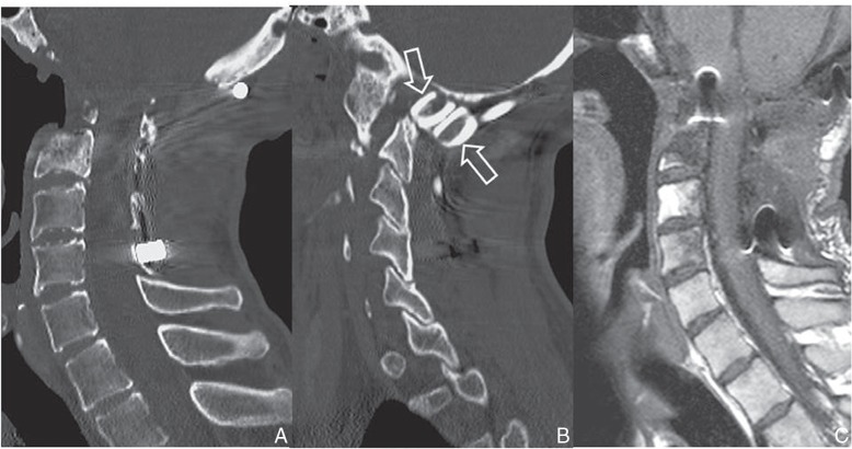
Fig. 6.
Two years after the reconstruction surgery, a mid-sagittal CT image of the upper cervical spine revealing odontoidectomy and C2–3 vertebrectomy defect (A). Parasagittal section showing autologous fibular grafts (open arrows), without resorption or bone fusion (B). Sagittal T1-weighted MRI showing sufficient decompression of the medulla oblongata and spinal cord (C). CT: computed tomography, MRI: magnetic resonance imaging.

