Fig. 3.

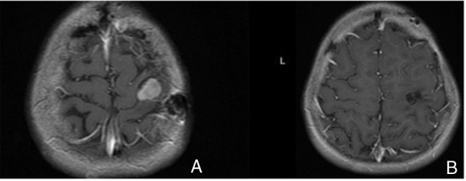
The lesion shows intense and homogeneous enhancement on postcontrast T1-weighted image (A) and gross-total removal of tumor (B).

The lesion shows intense and homogeneous enhancement on postcontrast T1-weighted image (A) and gross-total removal of tumor (B).