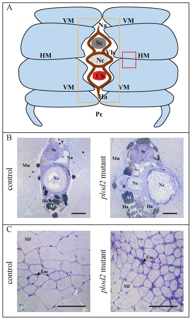
Figure 6. Histological analysis of the vertebral column of adult fish, cross sections.
The vertebral column of 4 months old adult fish was embedded, serially sectioned and stained with toluidine blue. A. Schematic representation of the transverse profile through the trunk of an adult zebrafish. Light blue segments represent the individual muscle segments that are separated from each other by sheets of matrix, namely the horizontal and vertical myosepta (HM, VM). The orange and red square indicate the region investigated in panel B and C respectively. B. Transverse sections at a position close to the intervertebral space (location of displayed sections is also indicated in figure 7A by asterisk), showing the presence of excessive bone (B) in Plod2 mutants. This bone prominently envelops the notochord (Nc), which is bent in the coronal plane (along the left-right axis), and is consequently sectioned twice in the same plane. C. Transverse sections demonstrating a reduced muscle fiber (Mf) diameter and the presence of a thicker endomysium (Em) between the muscle fibers, at the horizontal myoseptum of plod2 mutant fish. Other abbreviations: At, adipose tissue; Cb, caudal blood vessels; Ha, haemal arch; Mu, muscle tissue; Na, neural arch; Pc, peritoneal cavity; Sc, spinal cord; Vb, vertebral body. Scale bars represent 200 μm in B and C.

