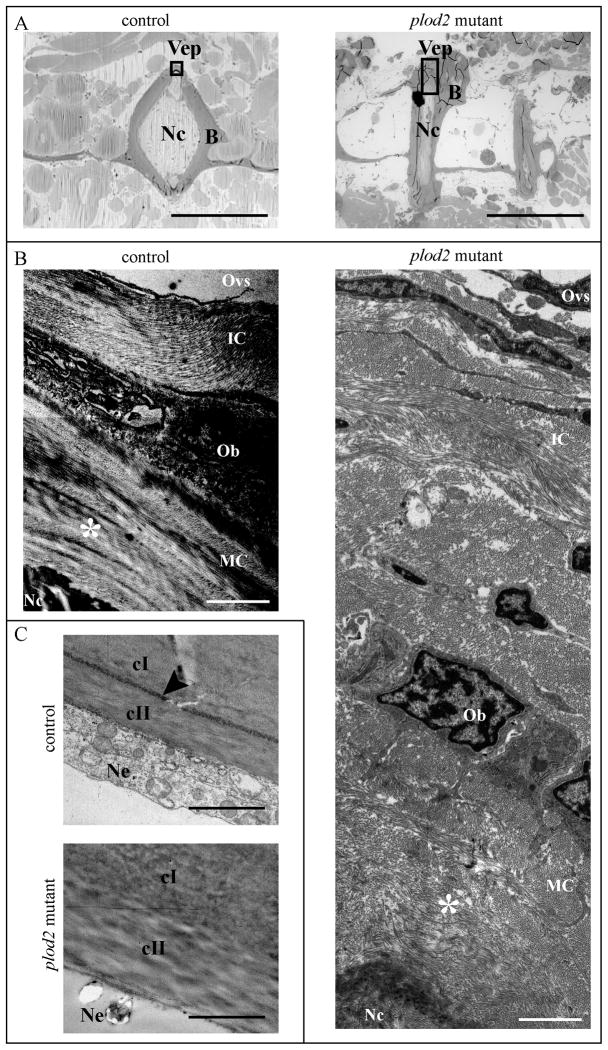
Figure 9. Analysis of bone type I collagen in adult fish using transmission electron microscopy (TEM).
A. Toluidine blue stained longitudinal sections of the vertebral column to illustrate where TEM images were acquired (black rectangles) both in control and plod2 mutant fish. B. TEM images of bone type I collagen in the vertebral end plates (Vep), which represents the growth zone of the vertebral bodies. Due to the presence of excessive periosteal bone (B) in mutant fish a larger image is shown for the plod2 mutant, compared to the control. Collagen is deposited by Osteoblasts (Ob) on the outer edges of the Vep as immature collagen (IC) and gradually matures towards the inside, adjoining the notochord (Nc), while developing a more organized structure. Hence mature collagen (MC) displays a typical plywood-like organization (indicated by asterisk), which is clearly visible in control fish but not in comparable regions (asterisk) for plod2 mutant fish. C. TEM images of the notochord sheath showing a region where the elastin layer delineating the notochord sheath (elastica externa, arrowhead), is clearly visible in control fish but interrupted in plod2 mutants. Also the collagen of the notochord sheath, which is mainly type II collagen (cII), has a disorganized appearance. Other abbreviations: cI, type I collagen; B, bone; Ne, notochord epithelium; Ovs, outer vertebral space; Vep, vertebral endplate. Scale bars represent 500 μm in A, 2 μm in B and 4 μm in C.

