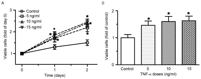
Figure 1.
TNF-α induced cell proliferation of RAECs. (A) Time-course and dose-response of RAEC proliferation following treatment with TNF-α. (B) Number of viable cells at 48 h following treatment with various concentrations of TNF-α. RAECs were seeded in a 96-well plate and were treated with various doses of TNF-α for 48 h; the number of viable cells was determined at the end of the experiment. Data are presented as the mean ± standard deviation of 6–10 individual samples of independent triplicate experiments. *P<0.05 vs. control group. RAECs, rat aortic endothelial cells; TNF-α, tumor necrosis factor-α.

