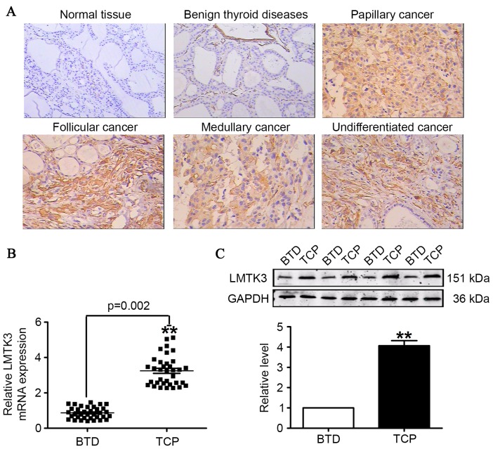
Figure 2.
LMTK3 level measured in TCP, BTD and HV tissue. (A) LMTK3 expression visualized by immunohistochemistry staining (original magnification, ×400; scale bar=100 µm). (B) The mRNA expression of LMTK3 was markedly elevated in TCP compared with BTD tissue samples. (C) Protein levels of LMTK3 were increased in TCP compared with BTD tissue samples. Data are expressed as the mean ± standard error of the mean (n=35). **P<0.01 vs. BTD group. LMTK3, lemur tyrosine kinase-3; GAPDH, glyceraldehyde 3-phosphate dehydrogenase; TCP, thyroid cancer patients; BTD, benign tumor diseases; HV, healthy volunteers.

