Figure 3.
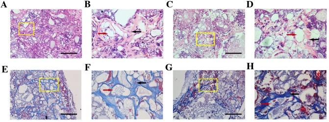
H&E and Masson staining of DKK3-shRNA DFCs and vector-infected DFCs. Representative images obtained following (A-D) H&E and (E-H) Masson staining. (A) A marked amount of mineralized matrices were deposited on the implant of DKK3-shRNA DFCs. Scale bar, 100 µm. (B) Local magnification of yellow box in A indicating limited space occurring between the HA/TCP (red arrow) and matrices. Collagen fibers and vasculogenesis (black arrow) are apparent. (C) Vector-infected DFCs presented the formation of numerous semi-bone matrices. Scale bar, 100 µm. (D) Local magnification of yellow box in C demonstrated that vasculogenesis and collagen fiber (black arrow) formation around the cells and HA/TCP (red arrow) were weak. (E) Newly formed blue osteoid matrices and collagen in DKK3-shRNA DFCs. Scale bar, 100 µm. (F) Local magnification of yellow box in E indicating mineralized matrices, collagen, newly formed vessels (black arrow) and HA/TCP (red arrow) constituted a network in the implants. (G) Vector-infected DFCs formed loose osteoid matrices and collagen. Scale bar, 100 µm. (H) Local magnification of yellow box in G indicating numerous intervals were formed in the osteoid matrix (black arrow) around HA/TCP (red arrow). H&E, hematoxylin and eosin; DKK3, Dickkopf-related protein 3; shRNA, short hairpin RNA; DFCs, dental follicle cells; HA/TCP, hydroxyapatite/β-tricalcium phosphate.

