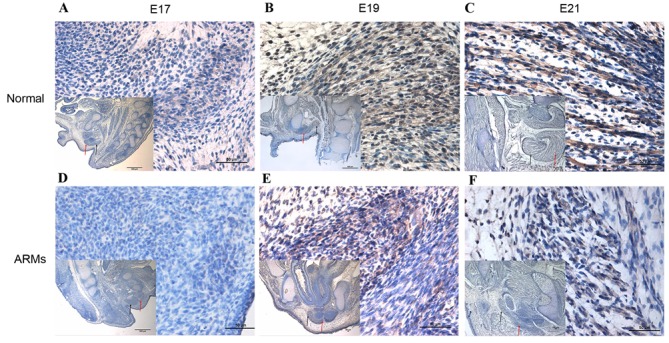
Figure 1.

Wnt3a expression in the SMC of normal and ARM model rats at various embryonic developmental stages. (A) At E17, immunoreactivity specific to Wnt3a was detected in the SMC. (B) At E19, positively-stained cells were mainly localized in the SMC and were observed in the bulbocavernosus muscle. (C) At E21, the Wnt3a immunoreactivity in SMC and bulbocavernosus muscle was higher than at earlier stages. (D) Sporadic positive staining cells could be detected in the SMC on E17. (E) At E19, the SMC were developed poorly, and marginal immunoreactivity specific to Wnt3a was detected in the SMC. (F) Wnt3a-positive cells increased slowly by E21 compared with earlier days. Magnification, ×400; magnification ×40 of figures in the lower left corner. The black arrows indicate SMC; the red arrows indicate the bulbocavernosus muscle. SMC, striated muscle complex; ARM, anorectal malformation; E, embryonic day.
