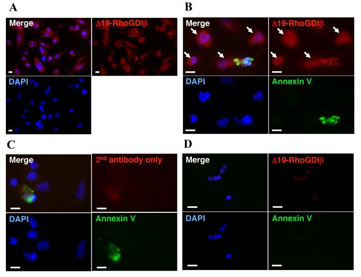
Figure 3.
Expression of Δ19-RhoGDIβ in non-apoptotic cells during the differentiation of THP-1 cells to macrophages. For immunostaining of differentiation-induced cells unattached dead and apoptotic cells were removed. The cells were cultured with 200 nM PMA for 3 days, and then cultured without PMA for a further 5 days. (A) Differentiated cells were stained with anti-Δ19-RhoGDIβ antibody (red). (B) Differentiated cells were stained with Annexin V (green) and anti-Δ19-RhoGDIβ antibody (red). Arrows indicate Δ19-RhoGDIβ-positive non-apoptotic cells. (C) Differentiated cells stained with Annexin V (green) and only with secondary antibody (red). (D) Unstimulated cells were stained with Annexin V (green) and anti-Δ19-RhoGDIβ antibody (red). Scale bar=20 µm. Similar results were obtained for three independent experiments. Representative results are shown. RhoGDIβ, Rho GDP-dissociation inhibitor β; PMA, PMA, phorbol 12-myristate 13-acetate; DAPI, 4′,6-diamidino-2-phenylindole.

