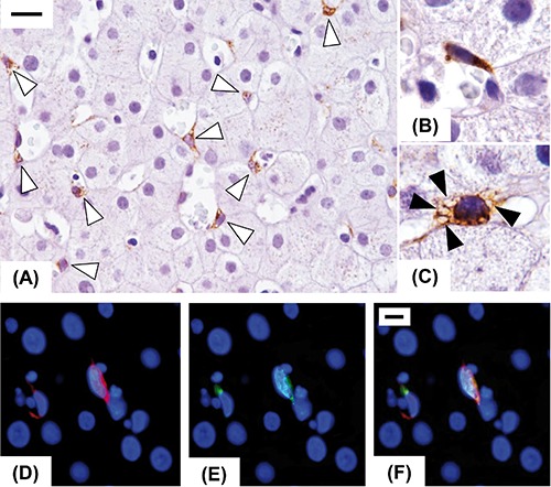
Figure 1.

Immunohistochemical expression of Reelin in injured human liver in early fibrosis stage. Reelin expression was restricted to spindle shaped cells (open arrowheads), resembling hepatic stellate cells, located in the perisinusoidal space (A). Some of them are elongated with a typical morphology of activated stellate cells (B), others exhibit a quiescent phenotype characterized by the presence of lipid droplets (closed arrowheads) in the cytoplasm (C). Double-labelling experiments with a hepatic stellate cells marker (CRBP-1, red, D) and Reelin (green, E), confirmed that Reelin was expressed by hepatic stellate cells (merge, F). Original magnification: A9), X400; B,C), X1000; D), X1000; scale bars: A), 20 µm; D), 10µm.
