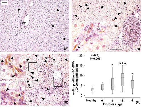
Figure 2.

Immunohistochemical expression of Reelin in healthy livers (A), livers with mild to moderate fibrosis (B) and with severe fibrosis (C). Reelin positive hepatic stellate cells/myofibroblasts (close arrowheads) are located mainly in the lobule and less frequently in the portal tract (PT) or septa (S). Reelin expression was also detected in cells lining putative lymphatic vessels (open arrowheads) and, faintly, in biliary ductules (*). The number of Reelin positive lobular HSCs/MFs increases with fibrosis progression (r=0.3, P<0.05; *P<0.05 vs healthy livers; #P<0.05 vs stage 0, ^P<0.05 vs stage 1, D). Original magnification: X200; high power fields: X400; scale bar: 50 µm.
