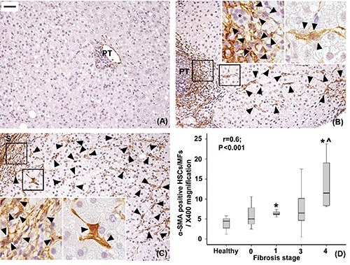
Figure 4.

Immunohistochemical expression of alpha-SMA in healthy livers (A), livers with mild-moderate fibrosis (B) and with severe fibrosis (C). Only very few alpha-SMA-positive hepatic stellate cells/myofibroblasts (arrowheads) are found in healthy livers and are individuated in the lobule, enlarged portal tract (PT) and septa (S) in fibrotic livers. The number of lobular alpha-SMA-positive hepatic stellate cells increases as fibrosis progresses (r=0.6, P<0.001; *P<0.01 vs healthy liver, ^P<0.01 vs stage 1, D). A control alpha-SMA-negative interlobular bile duct is indicated (*). Original magnification; X200; high power fields: X400; scale bar: 50 µm.
