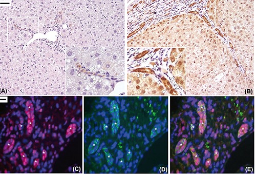
Figure 5.

Immunohistochemical expression of Dab1. In the healthy liver, only a faint expression of Dab1 was detected in a few ducts and ductules in the portal area (open arrowheads, A). Increased expression of disabled-1 protein was found in the biliary ducts and ductules of ductular reaction in the diseased liver with severe fibrosis (open arrowheads, B). Double-labelling experiments confirmed that Dab1 (red) was expressed at cytoplasmic and nuclear level by biliary cells of ductules of ductular reaction (asterisks) with the progenitor cell’s phenotype (EpCAM-positive cells, green) (C-E). Original magnification: X200 with high power fields X400 (A,B), X600 (C-E);scale bars: A,B), 50 µm; C-E), 10 µm.
