. 2016 Aug 9;8(1):325–333. doi: 10.1039/c6sc02411d

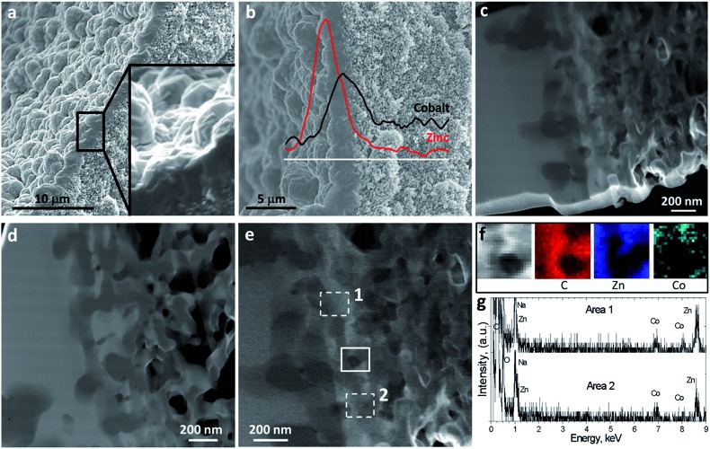
Fig. 3. Electron microscopy analyses of the double-layered ZIF-8/ZIF-9@P84 membrane. HF cross section depicts a 2.0 ± 0.4 μm thick ZIF layer together with a 3-fold magnification inset showing polyhedral ZIF crystals (a). The presence of outer-Zn from ZIF-8 and inner-Co from ZIF-9 coming from the stepwise microfluidic synthesis was confirmed by EDS (b). A wide MOF–support interphase was observed by STEM-HAADF in the lamella extracted by FIB, showing a strong ZIF–polymer interpenetration (c). HAADF Z-contrast shows the location of heavy Zn and Co atoms (lighter contrast) and polymer (dark contrast) (d). STEM-HAADF image magnification of the MOF–polymer interpenetration at a different location in the lamella (e). EDS elemental maps showing the spatial distribution of C, Zn and Co in the area marked with continuous line in (e) and (f). EDS analysis of areas 1 and 2 marked with dashed line in (e) and (g).

Fig. 3