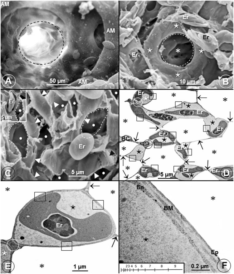
Fig 3. Infundibulae, air capillaries, blood capillaries and the blood-gas (tissue) barrier of the lung of the domestic fowl (Gallus gallus variant domesticus) and the Andean goose (Chloephaga melanoptera).
(A) A scanning electron micrograph showing an avascular infundibulum (dashed circle) of the lung of the domestic fowl. AM, atrial muscle. (B) Scanning electron micrograph of the lung of the Andean goose (Chloephaga melanoptera) showing an air capillary (* encircled by a dashed circle) which is surrounded by a single blood capillary (⋆). Er, erythrocytes. (C) Scanning electron micrograph showing lung parenchyma with extravasated erythrocytes (Er) that correspond in size (diameter) with those of the air capillaries (•). Some of the air capillaries are outlined with dashes lines. •, air capillaries; ►, blood capillaries. In the insert, an erythrocyte (Er) has slotted into an air capillary. (D, E) Low- (D) and high (E) magnification transmission electron micrographs showing air capillaries (*) and blood capillaries (⋆). In E, a blood capillary (⋆) is almost completely surrounded by air in the air capillaries (*). ↑, epithelial-epithelial cells connections; □, blood-gas (tissue) barrier; Er, erythrocytes; circle (O), blood capillary-blood capillary connection. (F) High magnification transmission electron micrograph showing the structure of the blood-gas (tissue) barrier. It [blood-gas (tissue) barrier] consists of an epithelial cell (Ep), an endothelial cell (En) and a basement membrane (BM). ⋆, blood capillary; *, air capillary. The insert shows a logarithmic scale that was used to determine the harmonic mean thickness of the blood-gas (tissue) barrier and that of the total barrier.

