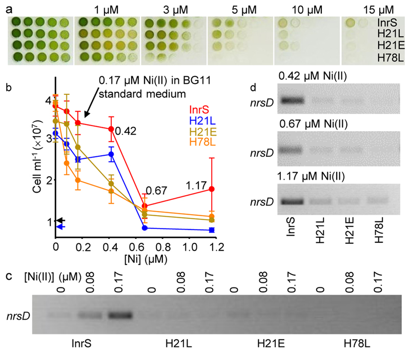
Figure 3. The tight Ni(II)-affinity of InrS was needed to regulate nrsD.
a. Spotting assay showing growth of strains InrS, H21L, H21E and H78L on standard BG11 medium plus the indicated [NiSO4]. b. Growth after 48 h in liquid medium (BG11 without NiSO4) plus the indicated [NiSO4]. Starting inoculum for H21L (blue) and other strains (black) indicated by arrows. Values are means of triplicate biological replicates with error bars representing ± one standard deviation. c. nrsD transcript abundance in media containing Ni(II) concentrations non-inhibitory to InrS cells (48 h) (loading controls shown in supplementary figure 4, full gel shown in supplementary figure 5). d. nrsD transcript abundance in media containing inhibitory Ni(II) concentrations (48 h) (all loading controls shown in supplementary figure 4, full gels shown in supplementary figures 6-8).

