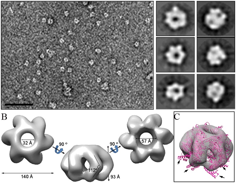
Fig 7. TdtA single-particle electron microscopy reconstruction.
(A) Representative electron micrograph of a negatively stained TdtA sample; bar = 50 nm. Six two-dimensional averaged classes of the oligomeric TdtA are shown (right). (B) Three-dimensional reconstruction of the hexameric TdtA. (C) Semitransparent model of the hexameric TdtA with the fitted atomic model of the hexameric HerA from S. solfataricus (pink). Arrows indicate the HerA region (residues 216–289), which remains outside the TdtA ring.

