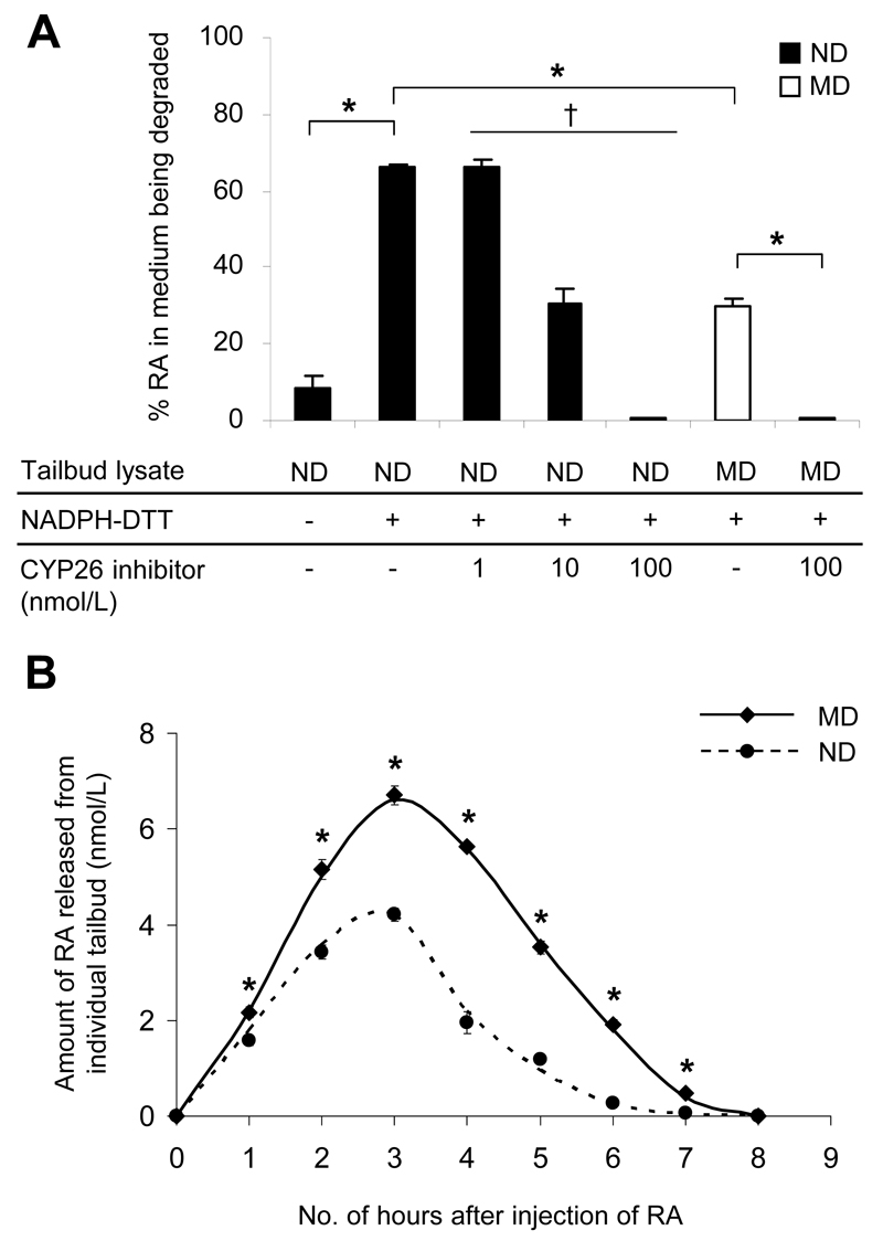
Figure 2.
Embryos of diabetic mice show reduced efficiency of RA catabolism. A: In vitro RA-degrading efficiency, presented as the percentage of RA in the medium being degraded by the tailbud lysate, in the presence or absence of the cofactor (NADPH) and the reducing agent (DTT) for optimal activity of CYP26 enzymes, and varying concentrations of R115866 (CYP26 inhibitor) (n = 18 in ND and MD groups with NADPH-DTT; n = 3-9 in other groups from 20 ND and 18 MD litters). *P < 0.001, Student t test; †R2= 0.742 and P = 0.001, linear regression. B: In vivo RA clearance measured as the amount of RA released from individual tailbuds, using a RA reporter cell line, at hourly intervals after injection of 50 mg/kg RA at E9 (n = 16-42 from three to eight litters). *P < 0.001 vs ND, Student t test and nonlinear regression. Error bars represent the mean ± SEM.

