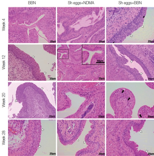
Fig. 3.

Tissue sections of mouse urinary bladder showing major histopathological abnormalities (HE stain). BBN group, hyperplasia at week 4, hyperplasia and dysplasia at week 12, 20, and 28 (white arrows); Sh eggs+NDMA group, normal at week 4 and squamous metaplasia at week 12, and dysplasia and hyperplasia at week 20 and 28 (white arrows); Sh eggs +BBN group, hyperplasia and dysplasia (white arrows) at week 4 and 12, enlarged and pleomorphic cells (black arrows) and epithelial vacuolar change and hyperplasia and dysplasia (white arrows) at week 20 and 28, respectively (original magnification, ×400).
