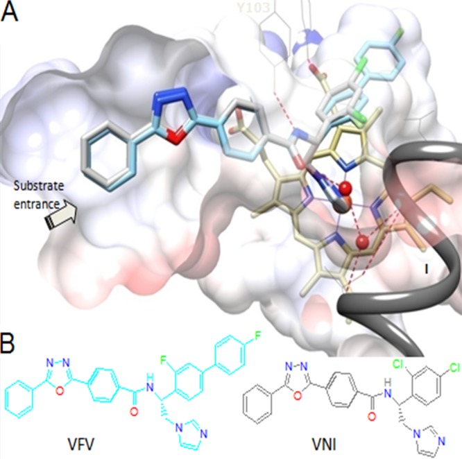
FIG 1.

(A) VNI (gray) and VFV (cyan) bound in the CYP51 active site (Protein Data Bank codes 3GW9 and 4G7G, respectively). Shown is a slice through the semitransparent protein surface, distal cytochrome P450 view. The heme is depicted in yellow; the catalytic iron atom is presented as an orange sphere. The H-bond network connecting the carboxamide fragment of the inhibitors with the CYP51 B′ helix (Y103) and helix I (proton delivery route) is displayed as dashed red lines, and water molecules are shown as red spheres. (B) Structural formulas. The color code for the atoms is the same as in panel A.
