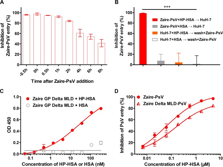
FIG 5.
HP-HSA inhibition of Zaire PsV entry at different intervals after addition of PsVs and its affinity of binding to purified Zaire GP trimer in vitro. (A) The inhibitory activity of 5 μM HP-HSA at 0, 0.5, 1, 2, 4, 6, and 8 h after Zaire PsV addition was compared with that of HP-HSA premixed with Zaire PsV 0.5 h before PsV addition. (B) HuH-7 cells were incubated with HP-HSA or HSA at 37°C for 1 h and washed with DMEM before Zaire PsV was added. As controls, HP-HSA or HSA was premixed with Zaire PsV before addition into wells plated with HuH-7 cells. (C) The binding affinities of Zaire GP (after the deletion of aa 314 to 462 and aa 637 to 676) and HP-HSA were demonstrated by using a binding ELISA and compared with the binding affinity of unmodified HSA. OD 450, optical density at 450 nm. (D) The entry-inhibitory activity of HP-HSA for Zaire PsV from which the GP MLD was deleted and wild-type Zaire PsV, which was utilized as a control. All experiments were performed in triplicate, and the error bars indicate standard deviations. The asterisks represent significant differences. ***, P < 0.001.

