FIG 4.
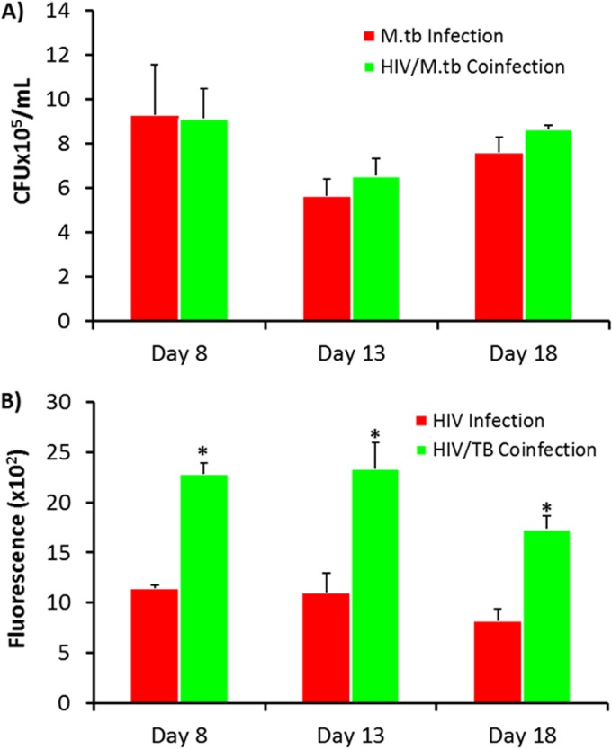
Quantitation of M. tuberculosis growth and HIV replication in MDMs coinfected with HIV-1 and M. tuberculosis. (A) M. tuberculosis growth was determined by counting of the number of CFU. (B) HIV-1 in human MDMs was quantitated by a reverse transcriptase assay. MDMs were infected with HIV-1 (MOI = 0.01) or M. tuberculosis (H37Ra; MOI = 1) or coinfected with HIV-1 and M. tuberculosis at days 8, 13, and 18 following differentiation. The coinfected MDMs and HIV-infected MDMs were further incubated for 11 days, and the medium was changed every 48 h. Supernatants from cells coinfected with HIV-1 and M. tuberculosis were harvested at day 11 for analysis of RT activity. Data represent the mean ± standard error of the mean for triplicate wells. Statistically significant differences were determined using Student's t test. *, P < 0.01 compared with HIV-infected cells in panel B.

