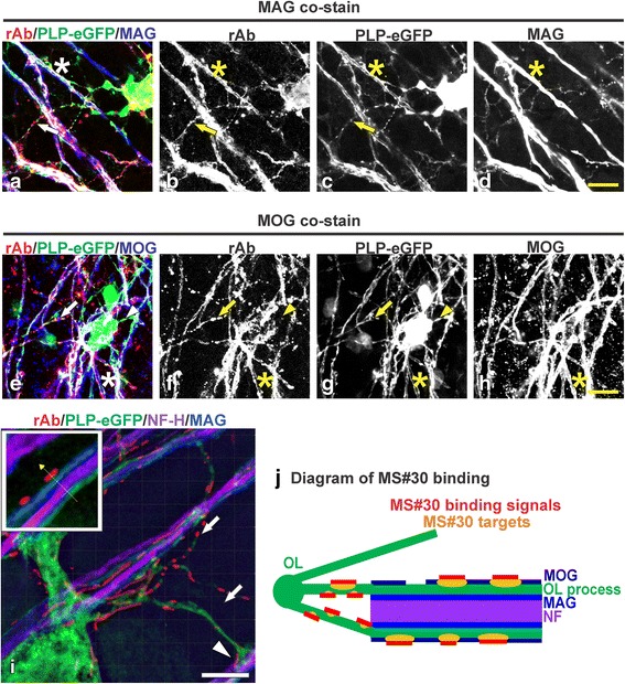
Fig. 1.

MS rAb #30 (MS#30) binds to oligodendrocyte processes and myelinated axons. Live cerebellar slices prepared from PLP-eGFP mouse pups were incubated with 20 μg/ml MS#30 rAb (red), then fixed and immunostained with anti-human IgG secondary and the indicated myelin markers. Confocal images of slices co-stained for the myelin proteins MAG (a–d; blue) and MOG (e–h; blue). MS#30 staining was co-localized with PLP-eGFP+ oligodendrocyte processes (arrows) and MAG+ or MOG+ myelinated axons (stars). Not all oligodendrocyte processes were stained with MS#30 (arrow heads). Scale bars: 10 μm. Super-resolution structured illumination microscopy image (i) of live slices stained with MS#30 (red), then fixed and stained for MAG (blue) and NF-H (purple). MS#30 reactivity was on oligodendrocyte processes (arrows), including those contacting adjacent axons (arrow head), and on myelinated MAG+ axons, outside of MAG layer (insert). Scale bar: 2 μm. Diagram of MS#30 rAb binding pattern (j)
