Fig. 3.

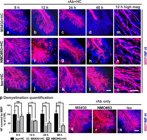
MS#30 and NMO rAb #53 (NMO#53) induce demyelination at different rates. Cerebellar slices were treated with MS#30 + HC (a–d, m), or NMO#53 + HC (e–h, n), or Iso + HC (i–l, o). At the indicated time points, slices were fixed and stained with MBP (red) and NF-H (blue) antibodies. Confocal images were taken with 25X (a–l) or 63X (m–o) objectives. The coverage of MBP on NF-H+ axons was quantified using a Matlab algorithm and normalized by control (Iso + HC treated slices) at 8 h (p). Statistical analyses were performed by multiple unpaired Student’s t test. *: p < 0.05, **: p < 0.01, ***: p < 0.001, ****: p < 0.0001, ns: not significant, n = 3-4. Confocal images of MBP and NF-H stained slices treated with rAbs alone at 48 h (q–s). Scale bars: 100 μm
