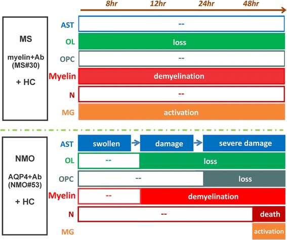
Fig. 8.

Schematic of distinct effects on glial and neuronal cells induced by MS and NMO antibodies in cerebellar slices. MS myelin-specific (myelin + Ab, such as MS#30 and MS#49) and NMO AQP4-specific (AQP4+ Ab, such as NMO#53) antibodies cause distinct patterns of complement-dependent tissue injury. Beginning at 8 h post-treatment, myelin-specific MS rAb MS#30 targets oligodendrocyte processes and induce rapid and increasing loss of oligodendrocytes, demyelination and microglia activation in the presence of complement (+HC). No significant effects on oligodendrocyte progenitors, neurons or astrocytes are observed. In contrast, NMO AQP4+ rAb targets astrocytes and induces robust complement-dependent astrocyte destruction. At 8 h post-treatment, astrocytes are swollen and the network of astrocytic processes is disrupted. At 48 h, massive destruction of the astrocyte network is apparent. Following astrocyte damage, there is a sequential loss of mature oligodendrocytes, demyelination, oligodendrocyte progenitor reduction, microglia activation and neuronal death [14]. AST: astrocyte, OL: oligodendrocyte, OPC: oligodendrocyte progenitor cell, N: neuron, MG: microglia
