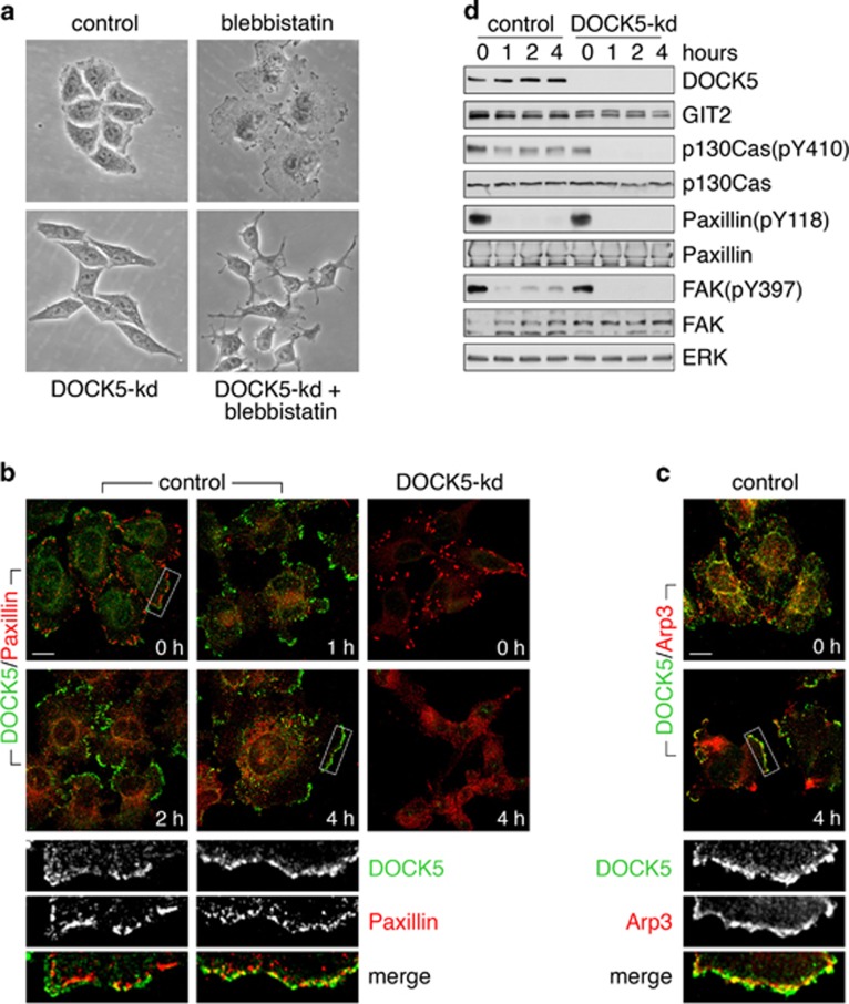
Figure 4.
Activity of DOCK5 is repressed by myosin II contractility. (a) HeLa cells were transfected with control or DOCK5 shRNA expression vectors and cultured in puromycin to select for expression of the resistance marker present in the vector. Cells were treated with the myosin II inhibitor blebbistatin (50 μM) or vehicle for 12 h and examined by phase contrast microscopy. (b) Localization of DOCK5 (green) and paxillin (red) in control and DOCK5-kd cells treated with blebbistatin for the indicated times. Scale bar indicates 10 μm. (c) Control cells treated with blebbistatin for 0 or 4 h were labeled to detect DOCK5 (green) and Arp3 (red). Scale bar represents 10 μm. (d) Western blot analysis of control and DOCK5-knockdown cells treated with blebbistatin for 0, 1, 2 and 4 h to detect phosphorylated forms and total levels of p130Cas, paxillin and FAK.

