Figure 8.
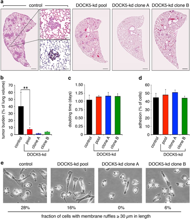
DOCK5-regulated metastasis of MDA-MB-231 is mediated through effects on motile/invasive capacities. (a) Representative images of tumor burden in lungs of nude mice 21 days after injection with control, DOCK5-kd pool, DOCK5-kd clone A and DOCK5-kd clone B cells. Scale bar represents 500 μm. The insets show adjacent sections of a small tumor stained with hematoxylin and eosin and human cytokeratin, respectively. Scale bar represents 50 μm. (b) Quantification of tumor burden in these mouse cohorts as described in Materials and methods (n=4 mice in each cohort). **P<0.02 by unpaired two-tailed Student's t-test. (c) Cell proliferation rates in control, DOCK5-kd pool, DOCK5-kd clone A, and DOCK5-kd clone B MDA-MB-231 cells (n=3). (d) Quantification of cell adhesion, as performed in Figure 1c using 40 μg/ml of collagen, in control, DOCK5-kd pool, DOCK5-kd clone A, and DOCK5-kd clone B MDA-MB-231 cells (n=3). (e) Morphology of control, DOCK5-kd pool, DOCK5-kd clone A and DOCK5-kd clone B MDA-MB-231 cells after overnight plating on collagen. The fraction of cells with membrane ruffles ⩾30 μm in length was quantified from 125 cells from each condition.

