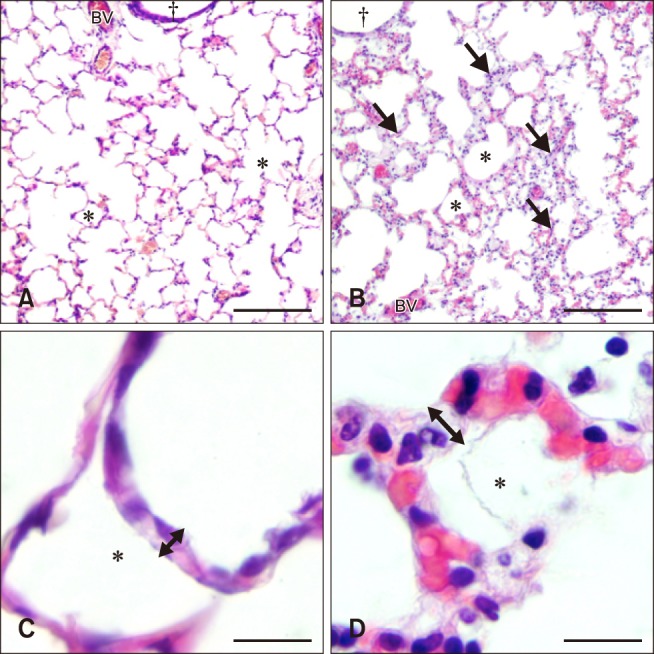
Fig. 1. Histology of hematoxylin and eosin stained lung sections from control (A and C) and septic (B and D) foals. Lung section from the control foal (A) shows normal lung morphology of bronchiole (†), blood vessels (BV), and alveolar space (*) compared to increased septal thickness and cellularity (arrows) in the lung section from the septic foal (B). Lung section from the control foal (C) shows normal alveolar septal width (double headed arrow) compared to the thickened septa of the septic foal lung section (D). Scale bars = 200 µm (A and B), 20 µm (C), 10 µm (D).

