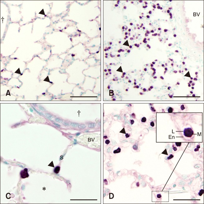
Fig. 3. Histologic lung sections from control (A and C) and septic (B and D) foals with MAC387 staining. Mononuclear cell (arrowheads) infiltration increased in septic (B and D) vs. control (A and C) foals. The inset image in figure d shows macrophage-like cells within the vasculature of the alveolar septa (S), suggesting the presence of a pulmonary intravascular macrophages. BV, blood vessel; En, endothelial cell; L, lumen; M, macrophage. *Alveolar space. †Bronchiole. Scale bars = 200 µm (A and B), 50 µm (C and D).

