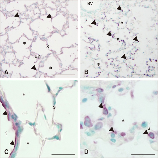
Fig. 5. Histologic lung sections from control (A and C) and septic (B and D) foals. Septic foals showed an increase in TLR9 staining (arrowheads) on mononuclear cells, but a slight decrease along bronchiole (†) epithelium, blood vessel (BV) endothelium, and alveolar septa (S). *Alveolar space. Scale bars = 100 µm (A and B), 50 µm (C and D).

