
Figure 3.
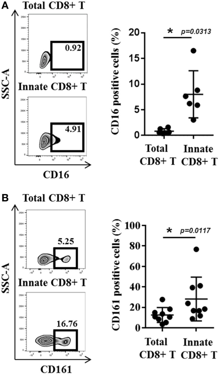
CD16 and CD161 are slightly enriched in innate CD8(+) T cells. (A) Peripheral blood mononuclear cells (PBMCs) from healthy donors (HDs) (n = 6) were cultured for 48 h in the presence of IL-15 and then analyzed by flow cytometry. One representative sample is shown for CD16 expression among total (upper panel) and innate (lower panel) CD8(+) T-cell subsets. Frequencies are displayed in the gate and the histogram (right panel) presents the full cohort of CD16-positive cells (mean ± SD) in both T-cell subsets. (B) PBMCs from HD (n = 9) were analyzed ex vivo by flow cytometry. One representative sample is shown for CD161 expression among total (upper panel) and innate (lower panel) CD8(+) T-cell subsets. Frequencies are displayed in the gates (left panel). The histogram (right panel) represents the full cohort of CD161-positive cells (mean ± SD) in both T-cell subsets.