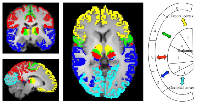
Figure 1.
Find-the-biggest segmentation of the thalamus into five clusters and their corresponding cortical target masks in a NS subject (x = 52, y = 48, z = 28). Occipital cluster 1 (light blue), temporal cluster 2 (dark blue), somatosensory postcentral cluster 3 (red), motor precentral cluster 4 (green), and frontal cluster 5 (yellow). The right part of the figure shows a color-coded schematic illustration of the thalamocortical connectivity.

