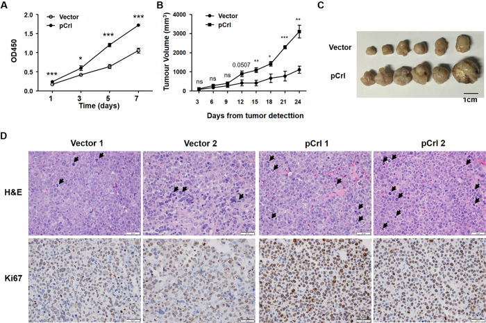
Figure 4. Reelin promotes MM cell growth in vivo.
(A) Reelin-over-expressing H929 cell lines proliferate faster than the controls. H929 cell lines with stable transfection of pCrl or control vector were seeded in FN-coated plates. The cell growth was analyzed by CCK8 method at 1, 3, 5, and 7 days later. A statistical significance (P < 0.0001) was found by the univariate analysis with Tukey post-hoc tests. The cell growth differences at the same time point were compared by Student’s t-test. (B,C) Reelin promotes MM tumor growth in vivo. Eight-week old female NOD/SCID mice were subcutaneously inoculated with vector- or pCrl-transfected H929 cell lines (1 × 10⁷) in 100 μL of serum-free RPMI-1640. Each group had 6 mice. The tumor size was measured in two perpendicular dimensions once every 3 days after palpable tumors developed (left panel). The mice were sacrificed at day 24 and the picture of tumors was taken (right panel). The Tukey post-hoc test was used to analyze the statistical significance between the two groups (P < 0.0001). The tumor size differences at the same time point were compared by Student’s t-test. (D) Reelin promotes MM cell proliferation in vivo. The tumor sections were evaluated by H&E and Ki67 staining. Arrowheads represent cells in cell cycle. Two representative tumor images from each group at 20x magnification were shown. The experiment was repeated twice and similar results were obtained.

