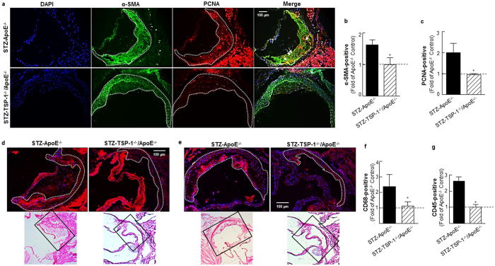
Figure 8. TSP-1 deficiency attenuates cell proliferation, lowers SMC abundance and reduces inflammatory cell content in aortic root lesions of hyperglycemic ApoE−/− mice.
Shown are representative images for (a) PCNA and α-SMA co-staining (10X magnification), (b,c) quantification data for PCNA and α-SMA staining (STZ-ApoE−/−: n = 7; STZ-TSP-1/ApoE: n = 6), (d) CD68 staining (15X magnification) depicting macrophage content and (e) CD45 staining (15X magnification) depicting leukocyte abundance and (f,g) summary data for quantification of CD68 (STZ-ApoE: n = 5; STZ-TSP-1/ApoE: n = 5) and CD45 (STZ-ApoE: n = 6; STZ-TSP-1/ApoE: n = 5) positive staining. Regions used for analysis are marked via dotted lines; arrows indicate PCNA-positive smooth muscle cells. Histology of the immunofluorescence images are shown in the corresponding H & E-stained images (indicated by black box). Results are presented as fold of ApoE−/− Control. All values are expressed are mean ± SD; *p ≤ 0.0019 vs. STZ-ApoE−/−.

