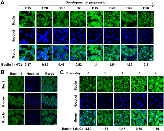
Figure 1. Beclin 1 is progressively relocalized to the nucleus during development and its nuclear distribution was reversed by starvation.
(A) Representative microscopic images of the subcellular localization of Beclin 1 in hepatic cells at different stages of mouse development are shown. Beclin 1 was labeled using DyLight 488 antibodies, and nuclei were stained using Hoechst 33342. The ratio between nuclear Beclin 1 (N) and cytoplasmic Beclin1 (C) was quantified in ≥50 cells using TCS-SP2 software and presented at the bottom of the images. (B) Representative microscopic images of the subcellular localization of Beclin 1 in the heart, kidney, and muscle tissues from adult C57BL/6J mice (6–8 weeks old). (C) Representative microscopic images of Beclin 1 subcellular localization in adult mouse hepatocytes during 4 days of starvation (Starv). The ratio of nuclear Beclin 1 (N) and cytoplasmic Beclin1 (C) was presented at the bottom of the images. Similar results were observed in at least three independent experiments. Scale bar, 20 μM.

