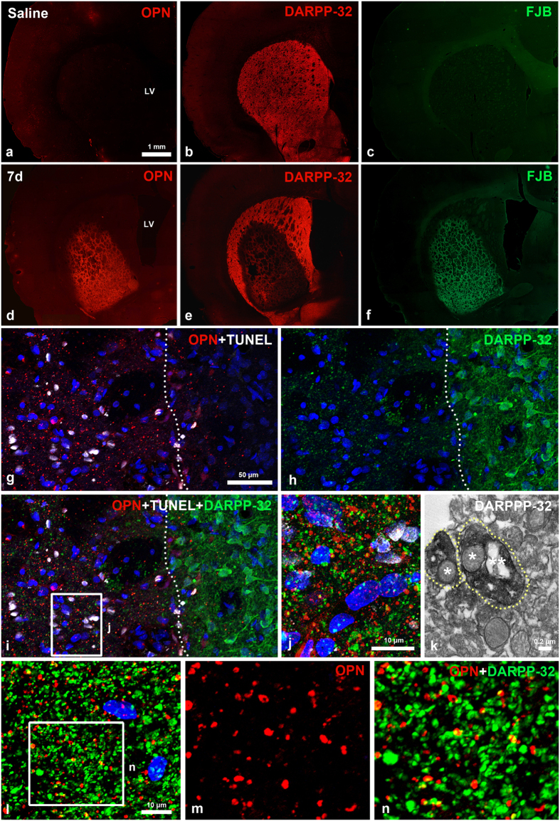
Figure 2. Spatial relationships between OPN and neuronal cell injury induced by 3-NP.
(a–f) Serial sections of 3-NP-treated rat forebrain 7 days post-lesion are stained with OPN and dopamine- and cyclic AMP-regulated phosphoprotein with an apparent molecular weight of 32 kDa (DARPP-32; red channel), and Fluoro-Jade B (FJB; green channel). Note that a prominent OPN expression is induced within the lateral striatum, in which concomitant loss of DARPP32-positive neurons and intense FJB staining are visible in rats intoxicated with 3-NP. (g–i) Triple labelling for OPN, terminal deoxynucleotidyl transferase dUTP nick end labelling (TUNEL) and DARPP-32. OPN and TUNEL staining are restricted to the lesion core (left side of white broken line), whereas DARPP-32-positive neuronal soma with processes are evident in the perilesional area. (j) Higher magnification view of the lesion core (boxed area in i) showing the punctate distribution of OPN- and DARPP-32-positive profiles. (k) Ultrastructural features of DARPP-32-positive profiles. Dotted DARPP-32 corresponds to the immunolabelled dendrites (delimited by white dotted lines) containing the degenerating (one star) and normal-appearing (two stars) mitochondria. (l–n) Double-labelling for OPN and DARPP-32 showing that most of OPN-positive dots are co-labelled with DARPP-32, but some are devoid of DARPP32 staining, and vice versa. (m, n) Higher magnification views of the boxed area shown in i. Cell nuclei appear blue after DAPI staining.

