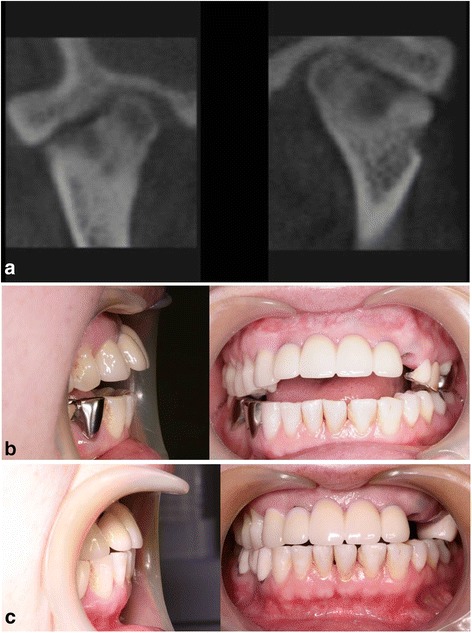
Fig. 3.

Case 3. a CT images showing flattening of bilateral condylar heads, irregularity of articular surface, and narrowing of joint spaces. b Anterior open bite developed with overbite changing from 0 to −6 mm during splint therapy. c Anterior open bite was closed, and no further progression of anterior open bite has been observed
