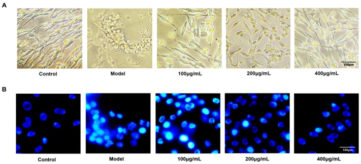
Figure 2.
Morphology and fluorescent chemical staining of PC12 cells. (A) The changes of morphology of PC12 cells in different groups under light microscope observation. Fucoidan attenuated the morphological changes induced by Aβ + d-Gal, which resulted in decreased and shortened nerve filaments and cell aggregation. (B) Hoechst 33258 staining of PC12 cells injured by Aβ and d-Gal with different concentration of fucoidan (100, 200, 400 μg/mL). Magnification scale is 400×.

