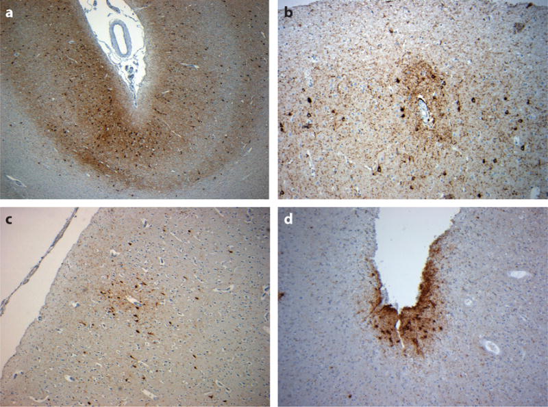
Figure 2.

Neocortical tau pathology in chronic traumatic encephalopathy. Tau immunoreactive profiles are distributed throughout the neocortex, although they typically show a preferential distribution toward the superficial neocortical layers and depths of sulci [a, 49-year-old male 12 months following single, severe traumatic brain injury (TBI)], with a distinctive and characteristic perivascular accentuation of immunoreactive neurons and glia, whether exposed to repetitive, mild TBI (b, 56-year-old male, former rugby player) or a single, moderate or severe TBI (c, 48-year-old male 3 years following a single, severe TBI). The accumulations of subpial thorn-shaped astrocytes may also be observed (d, 59-year-old male, former soccer player). All sections stained for phosphorylated tau using antibody CP-13 (courtesy of Dr. P. Davies).
