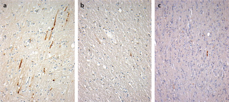
Figure 4.

Axonal pathology in the corpus callosum with varying durations of survival after traumatic brain injury (TBI). A constant in all severities of TBI is diffuse axonal injury (DAI) with associated interruption of axonal transport. In tissue sections, DAI is typically revealed by staining for amyloid precursor protein (APP) and is detectable in under an hour following injury as immunoreactive axonal profiles with varying abnormal morphologies (a, 18-year-old male 11 h after severe TBI). Beyond this acute-phase axonal injury, evidence of ongoing interruption of axonal transport is marked by scattered, morphologically abnormal axons; staining for APP remains present in survivors 1 year or more after a single, moderate or severe TBI (b, 24-year-old male 8 years after a single, severe TBI) and in material from individuals exposed to repetitive, mild TBI (c, 59-year-old male, former soccer player). All sections stained for an antibody to the N-terminal amino acids 66–81 of APP (EMD Millipore).
