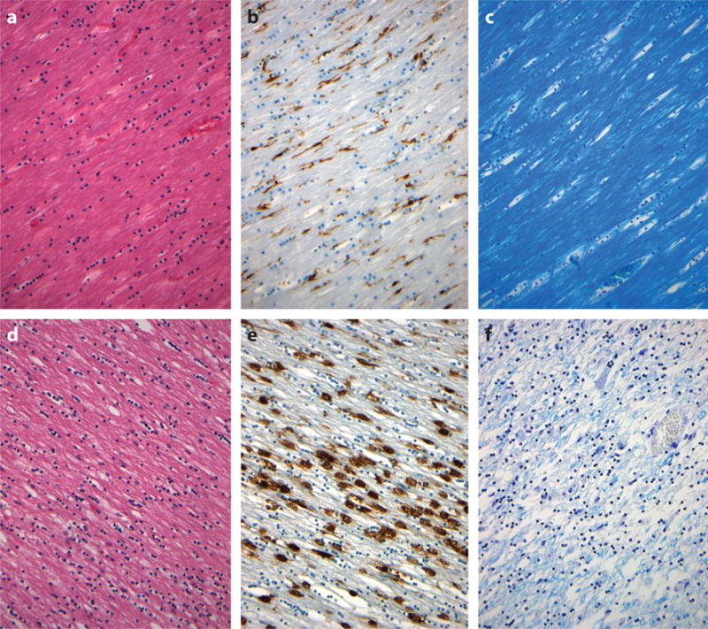
Figure 6.

Neuroinflammation and degradation of white matter in the corpus callosum occurring with survival following traumatic brain injury (TBI). Material from approximately 30% of survivors at 1 year or more after a single, moderate or severe TBI shows evidence of white matter degradation as rarefaction in staining with Hematoxylin and eosin (d) when compared with material from uninjured controls (a). Accompanying this is evidence of ongoing neuroinflammation in the form of numerous amoeboid, activated microglia (e) in contrast to the quiescent, ramified microglia in uninjured controls (b). Staining for myelin with Luxol fast blue–Cresyl violet demonstrates an associated loss of myelin with evidence of continued myelin degradation after trauma (f). (a–c) Sections from the corpus callosum of a 38-year-old male non-TBI control whose cause of death was sudden, unexpected death in epilepsy. (d–f) Sections from the corpus callosum of a 56-year-old male with 3-year survival after a single, severe TBI. (b) and (e) stained for HLA-DP, -DQ, and -DR using antibody CR3/43 (Dako, Agilent Technologies) to reveal activated microglia.
