
Figure 7.
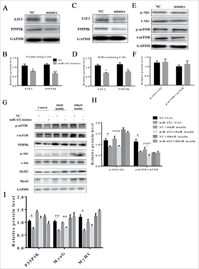
MiR-432 blocked myogenesis through PI3K/AKT signaling pathway. (A) Western blot analysis of protein change of E2F3 and P55PIK during proliferation. (B) Quantification of E2F3 and P55PIK protein expression in proliferating cells. (C) Protein changes of E2F3 and P55PIK during myoblast differentiation. (D) Quantification of E2F3 and P55PIK protein expression in differentiating cells. (E) Western blot analysis of signal molecules of PI3K/Akt/mTOR pathway during proliferation. (F) Ratios of p-Akt/t-Akt and p-mTOR/t-mTOR in proliferating cells. (G) Protein level of signal molecules in PI3K/Akt/mTOR pathway. After transfection with NC or miR-432 mimics, myoblasts were induced by myogenic differentiation medium for 3 days followed by incubating with control, 10nM insulin or 100nM insulin respectively for 24 h. Total protein was collected for western blot analysis. (H) The ratios of p-Akt/t-Akt and p-mTOR/t-mTOR protein changes in 7G. (I) Quantification of P55PIK, MyoG, and MyHC protein changes in 7G. Data were shown by mean ± SD of three independent experiments. *, P < 0.05;**, P < 0.01.