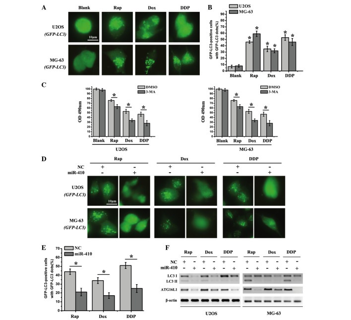
Figure 4.
miR-410 effectively inhibits autophagy in osteosarcoma cell lines. (A) U2OS-GFP-LC3/MG-63-GFP-LC3 cells which stably express GFP-LC3 were used to detect autophagy after the cells were treated with various agents for 48 h (Rap, 20 nM; Dox, 0.1 µg/ml; DDP, 10 µM). (B) Percentage of GFP-LC3 punctate-positive cells was quantified and analyzed by automated image acquisition using a threshold of ≥5 dots/cell. (C) Cell viability was detected after the cells were treated with various agents for 48 h with the presence or absence of the autophagy inhibitor 3-MA. (D) U2OS-GFP-LC3/MG-63-GFP-LC3 cells which stably express GFP-LC3 were used to detect autophagy after the cells were treated with Rap (20 nM), Dox (0.1 µg/ml) or DDP (10 µM), and were transfected with miR-410 or NC mimics for 48 h. (E) Percentage of GFP-LC3 punctate-positive cells was quantified and analyzed by automated image acquisition using a threshold of ≥5 dots/cell (U2OS-GFP-LC3). (F) Western blot analysis of LC3I/II and ATG16L1 expression from the lysates of U2OS and MG-63 cells transfected with NC or miR-410 for 48 h. β-actin was used as an internal control. Data are presented as the mean ± standard deviation and are representative of three independent experiments (*P<0.05). ATG161L, autophagy related 16-like 1; miR-410, microRNA-410; NC, negative control; Rap, rapamycin; Dox, doxorubicin; DDP, cisplatin; GFP, green fluorescent protein; LC3I/II, microtubule-associated protein 1A/1B-light chain 3; OD, optical density; 3-MA, 3-methyladenine; DMSO, dimethyl sulfoxide.

