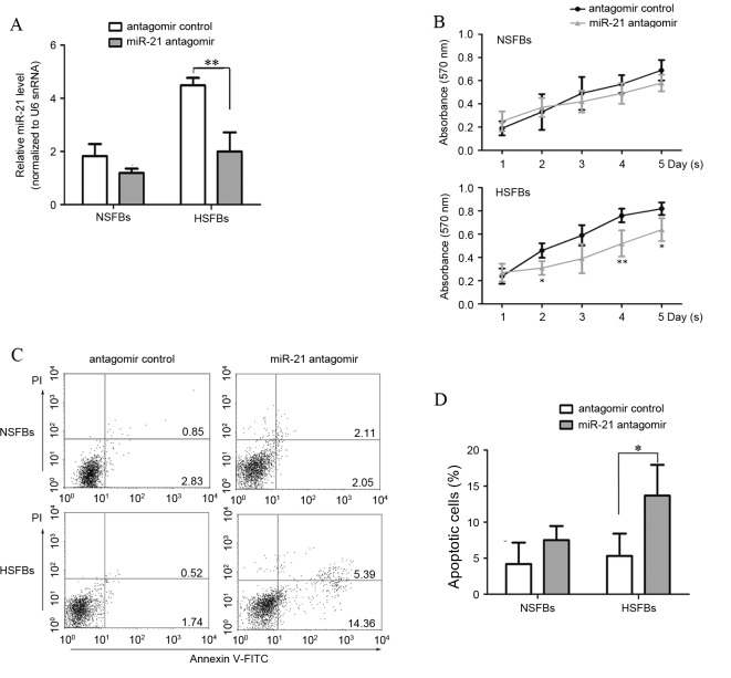
Figure 3.
miR-21 antagomir inhibits proliferation and induces apoptosis in HSFBs. HSFBs and NSFBs were treated with miR-21 antagomir or miR-21 antagomir control for 48 h. (A) At 48 h after transfection, miR-21 expression levels were evaluated by reverse transcription-quantitative polymerase chain reaction analysis. (B) MTT assays were performed at 24 h intervals for five days. *P<0.05, **P<0.01 vs. the antagomir control. (C) Apoptosis was measured by flow cytometric analysis using Annexin V and PI staining. Right lower quadrant, early apoptotic cells; right upper quadrant, late apoptotic cells. (D) The rate of apoptosis in different groups was calculated and compared. miR, microRNA; HSFBs, hypertrophic scar fibroblasts; NSFBs, normal skin fibroblasts; PI, propidium iodide; snRNA, small nuclear RNA. *P<0.05, **P<0.01.

