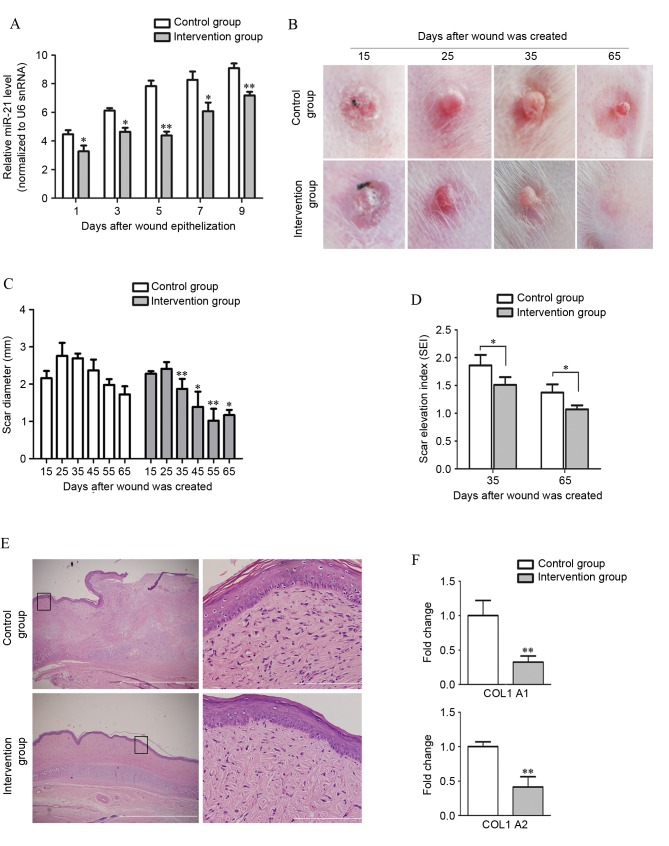
Figure 5.
Local treatment with miR-21 antagomir significantly decreases scar formation. (A) miR-21 antagomir administration in the intervention group significantly suppressed the upregulation of miR-21 in scar tissue (n=3) following wound epithelialization, as compared with the miRNA antagomir control administration group. (B) Representative images of scar formation in the rabbit ear model. Scars in the group withanti-miR-21 intervention exhibited a delay in formation process compared with the control group. (C) Quantification of formed HS. The diameter of scars in both groups was measured with a slide gauge. Between day 25 and day 65, the diameter of scars in the miR-21 antagomir intervention group was significantly smaller compared with the control group. (D) Comparison of SEI on days 35 and 65. The degree of dermal hypertrophy of each scar was expressed as the SEI. This index is the ratio of the area of newly formed dermis of the scar to the area of surrounding normal dermis. (E) Pathological analyses by hematoxylin and eosin staining confirmed that scar formation was inhibited in the miR-21 antagomir-treated group when comparing the collagen distribution on day 65. The distribution of collagen fibers in the control group were dense, enlarged and disordered, with more cells in the epidermis and dermis of HS, while collagen fibers were more mature and well arranged with few cells in the intervention group. Boxes in the left column indicate the areas that are enlarged in the corresponding panels in the right column. Scale bars: 1 µm (right) and 50 µm (left). (F) Expression of COL1A1 and COL1A2 in different groups of scars was measured and compared by reverse transcription-quantitative polymerase chain reaction analysis. miR, microRNA; miRNA, microRNA; HS, hypertrophic scar; SEI, scar elevation index; snRNA, small nuclear RNA; COL1A1, collagen type I α 1 chain; COL1A2, collagen type I α 2 chain. *P<0.05, **P<0.01 vs. the control group.

