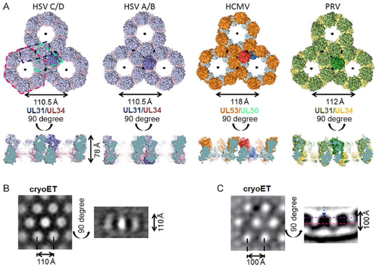
Fig. 5.

Comparison of hexagonal lattice in HSV-1 (two crystal lattices), HCMV (crystal lattice), and PRV (model derived from cryoET data). (A) For each lattice, three connected hexameric rings are shown side by side in a top view, perpendicular to the sixfold symmetry axis, and a side view. One NEC heterodimer is highlighted in every lattice. The two-, three-, and sixfold axes in each lattice are indicated by lense, triangle, and star symbols, respectively. Representative dimer, trimer, and hexamer are indicated by dashed lines in the HSV-1 C/D lattice. The hexameric rings are very similar in both HSV-1 lattices and the HCMV lattice but differ in the PRV lattice model. HSV-1 A/B is rings are turned toward each other in a 10.5 degree angle compared to the C/D lattice. The PRV lattice model is the only curved lattice in the side view while the rest of the lattices are planar. All crystal lattices are ∼78 Å thick. For HSV-1 NEC (B) and PRV NEC (C), cryoET lattices are shown for comparison. The lattices are thicker than in the crystal structures due to the presence of the membrane-proximal regions, absent from all crystallized NEC constructs. The diameter of the hexameric ring in the PRV cryoET lattice is slightly smaller than other ring diameters due to the curvature of the lattice and the positioning of the slice (purple dashed line in side view). HSV-1 NEC cryoET image in (B) is reprinted from Bigalke, J.M., Heldwein, E.E., 2015b. Structural basis of membrane budding by the nuclear egress complex of herpesviruses. EMBO J., 34, 2921–2936. PRV NEC cryoET image in (C) is reprinted from Hagen, C., Dent, K.C., Zeev-Ben-Mordehai, T, Grange, M., Bosse, J.B., Whittle, C., Klupp, B.G., Siebert, C.A., Vasishtan, D., Bauerlein, F.J., Cheleski, J., Werner, S., Guttmann, P., Rehbein, S., Henzler, K., Demmerle, J., Adler, B., Koszinowski, U., Schermelleh, L., Schneider, G., Enquist, L.W., Plitzko, J.M., Mettenleiter, T.C., Grunewald, K., 2015. Structural basis of vesicle formation at the inner nuclear membrane. Cell, 163, 1692–1701. http://dx.doi.org/10.1016/j.cell.2015.11.029, under Creative Commons Attribution License, https://creativecommons.org/licenses/by/4.0/.
