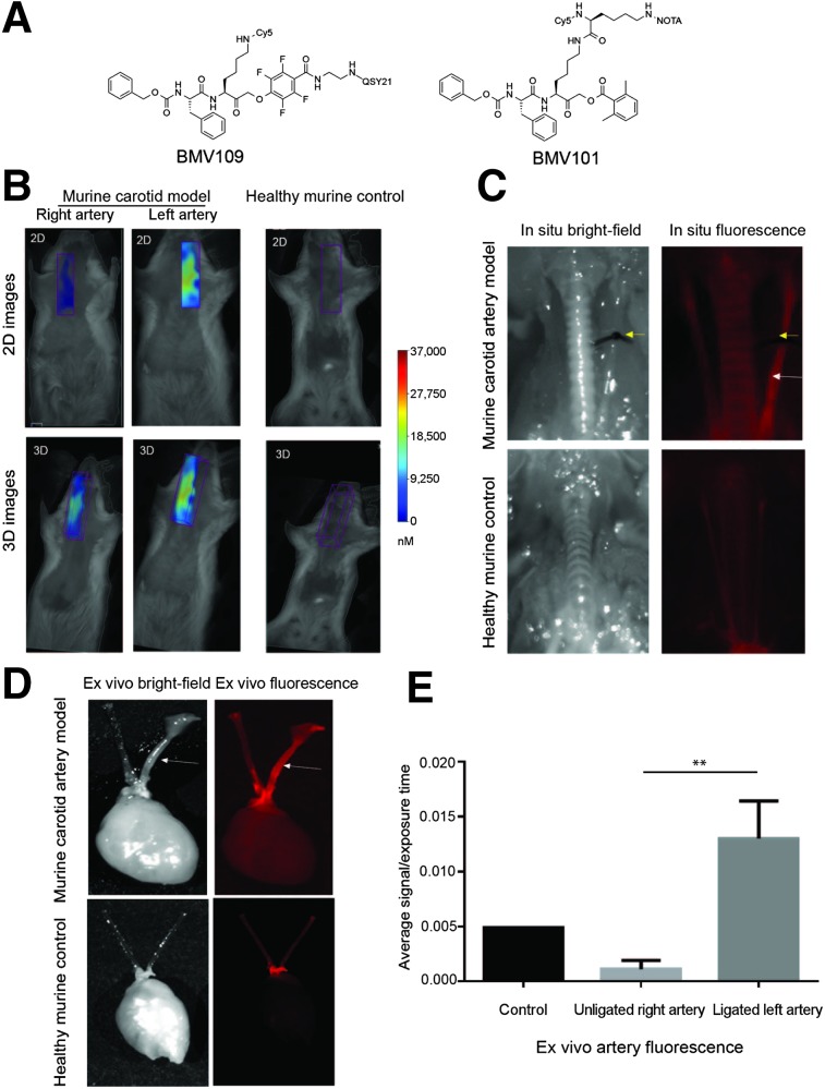
FIGURE 1.
Application of activity-based probe BMV109 in experimental carotid inflammation model. (A) Structure of fluorescent cathepsin probe BMV109. (B) Noninvasive FMT imaging in murine carotid arteries and healthy control arteries. Both 2- and 3-dimensional images show high signal in left carotid artery of diseased mouse compared with right artery and control. (C) Corresponding in situ fluorescence imaging of BMV109 in murine carotid arteries and control healthy mouse. (D) Ex vivo florescence imagining of diseased and healthy carotid arteries. (E) Quantitative analysis of ex vivo fluorescence showed significantly higher signal in left ligated carotid artery than the nonligated carotid artery and control. n = 3, **P < 0.005 by t test.

